Cluster Experiments at the Free-Electron-Laser FLASH
Highly intense radiation in the XUV-regime at the Free-Electron-Laser FLASH at DESY is used to investigate structures and properties of metal clusters by means of photoelectron spectroscopy and diffractive imaging. By XUV-photoemission, the complete electronic valence band and low-lying core-levels become accessible. Single-shot scattering experiments with XUV-light reveal geometric structures of metal clusters, including information on shape and orientation at the instance of the femtosecond XUV-pulse.
Core-Level Spectroscopy of Metal Clusters
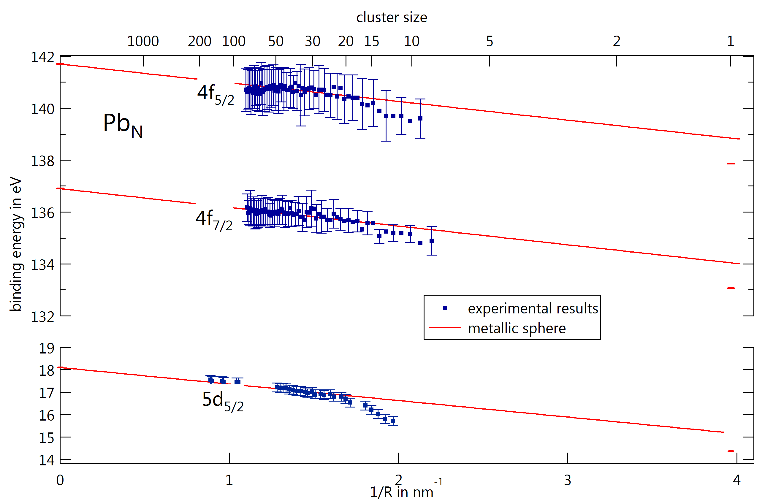
As known for valence levels, also shallow core-levels of metal clusters show an energy shift as a function of cluster size. For large cluster (typically more than hundred atoms), these shifts are explained by the metallic sphere model, i.e. the clusters show metallic character. However, as the cluster size decreases, the energy shift of the core-levels deviates from the metallic sphere model, as has been shown for lead clusters with less than 20 atoms. This changing behavior indicates a reduced effect of core-hole screening by the electrons, i.e. the electronic structure loses its metallic character.
Core-Hole Screening as a Probe for a Metal-to-Nonmetal Transition in Lead Clusters
V. Senz et al., Phys. Rev. Lett.102, 138303 (2009)
Pb 4f photoelectron spectroscopy on mass-selected anionic lead clusters at FLASH
J. Bahn et al., New J. Phys.14, 075008 (2012)
The experiments are realized by a collaboration of several universities. The project is funded by the Federal Ministry of Education and Research, bmbf. Together with the project partner at the University of Freiburg, we currently design and realize a new and versatile electron-spectrometer system for studies of free atomic nanoparticles at free-electron lasers, like FLASH.
project leaders
- Dr. Josef Tiggesbäumker, Prof. Dr. Karl-Heinz Meiwes-Broer, Universität Rostock
- Prof. Dr. Bernd v. Issendorff, Universität Freiburg
collaborators
- Prof. Dr. Thomas Möller, Technische Universität Berlin
- Prof. Dr. Gerd Ganteför, Universität Konstanz
- Prof. Dr. Eckart Rühl, Freie Universität Berlin
Metal Cluster Imaging
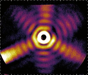
Wide-angle scattering of the femtosecond short XUV-pulses of FLASH at free particles of several nanometers in size is used to obtain single-shot images of individual metal clusters. From the scattering images information on instantaneous cluster shape and orientation are deduced by a dedicated data evaluation.
The 3D-architecture of individual free silver nanoparticles captured by X-ray scattering
I. Barke et al., Nature Communications 6, 6187 (2015)
The project is a collaboration of the TU Berlin, Uni Rostock, Uni Freiburg, and DESY (FLASH)
- Dr. Daniela Rupp, Technische Universität Berlin
- Dr. Ingo Barke, Universität Rostock
- Prof. Dr. Thomas Möller, Technische Universität Berlin
- Prof. Dr. Bernd von Issendorff, Universität Freiburg
- Prof. Dr. Thomas Fennel, Universität Rostock
- Prof. Dr. Karl-Heinz Meiwes-Broer, Universität Rostock